Congenital Heart Disease

Congenital heart disease (CHD) refers to a structural abnormality of the heart that is present from birth. These abnormalities develop due to abnormal development of the fetus within the uterus. Congenital heart diseases are the most common birth defects in new-born babies and 9 out of every 1000 babies have a congenital heart defect.
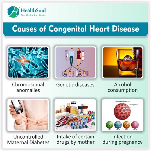
Causes of CHD
There are numerous causes that can lead to the development of an abnormal heart in the baby. These are as follows:
- Chromosomal anomalies: A decrease or excess of chromosomes can lead to many diseases that are frequently associated with CHD. Down Syndrome (extra chromosome 21) and Turner Syndrome (absence of one X chromosome) are common diseases with associated CHD.
- Genetic diseases: Presence of abnormal genes can also lead to defects
- Maternal smoking or alcohol consumption: Cigarette smoke and alcohol are known teratogens (substances with potential to cause congenital defects) and can often lead to defects in the heart.
- Uncontrolled Maternal Diabetes: High blood sugar during critical times of fetal development can lead to abnormalities in the heart
- Intake of certain drugs by the mother: Some medications like lithium, thalidomide, ACE inhibitors and isotretinoin (for acne) are all known to cause CHD
- Infections during pregnancy: Rubella is an important infection which if it occurs during early pregnancy can cause congenital rubella syndrome, marked by patent ductus arteriosus.
Types of CHD
- Presence of a Hole in the heart: These defects are marked by mixing of oxygen rich blood on the left side of the heart with oxygen depleted blood on the right side of the heart. This results in supply of blood with low oxygen content to the body.
- Ventricular Septal Defect (VSD): There is a hole or defect present in the septum dividing the left and right ventricles of the heart. A VSD may be associated with other heart defects or may be the only lesion. Some VSDs may be very small and close spontaneously in childhood whereas others may be very large and present early in life with rapid progression of symptoms.
- Atrial Septal Defect (ASD): This occurs due to a defect in the septum dividing the left and right atria. Often the individual is asymptomatic till the 3rd to 4th decade when they start to experience shortness of breath requiring evaluation and care.
- Patent Foramen Ovale (PFO): Prior to birth, there exists a communication between the left and right atria known as the foramen Ovale. This is guarded by a flap and is necessary for normal circulation in the foetus. After birth, it closes spontaneously. Failure to close completely leads to PFO. A PFO is considered to be a normal variant and does not cause any symptoms.It is important to note however, that a PFO can allow a clot forming in the body, to move from the right to the left side of the heart and reach the brain to cause a stroke.
- Patent Ductus Arteriosus (PDA): The ductus arteriosus is a connection between the aorta (carrying oxygen rich blood to the body) and the pulmonary artery (carrying oxygen depleted blood to the lungs). This is a normal and necessary connection in the fetus which slowly closes after birth. Failure to close within a couple of days following birth is abnormal and leads to mixing of blood with poor supply of oxygen rich blood to the body.
- Abnormal Blood Vessels: Abnormal positions and structure of the major blood vessels can lead to problems after birth.
- Transposition of the great arteries: This occurs when the aorta and the pulmonary artery are interchanged in position. This causes 2 closed circuits in the baby rapidly leading to death if not addressed in the first week of life. Generally an associated VSD or PDA is present to allow some degree of mixing of blood between the two circuits.
- Coarctation of the Aorta: This is a narrowing of the aorta leading to a reduction in blood flow to the body parts present beyond the obstruction. This is often not detected till later in life.
- Total Anomalous Pulmonary Venous Connection: This is a rare condition when the pulmonary veins (carrying oxygen rich blood from the lungs to the heart) attach to the wrong side of the heart
- Heart Valve Abnormalities: There are 4 heart valves and abnormalities can develop in all of the them. Thickening of the valve can obstruct blood flow leading to stenosis and weakening can lead to leakage causing regurgitation. The most common is the bicuspid Aortic Valve
- Bicuspid Aortic Valve: This is the most common CHD. There are 2 cusps in the valve instead of the normal 3. This doesn’t cause any problems early in life apart from an abnormal heart sound. However, with age, a bicuspid valve is at greater risk for calcification and can cause obstruction to blood flow leading to aortic stenosis and also weaken, leading to leakage and aortic regurgitation.
- Combination of several defects: Several different types of developmental abnormalities can be present in the heart. The most common being Tetralogy of Fallot.
- Tetralogy of Fallot (TOF): This CHD comprises of 4 abnormalities- Pulmonary stenosis (thickening of pulmonary valve), VSD, Overriding Aorta (Aorta sits on the ventricular septum), and hypertrophic right ventricle (thickened right ventricle muscle). Depending on the severity of the defects, the age at presentation may vary.

Symptoms of CHD
- Shortness of breath or rapid breathing in infants
- Cyanosis or blue discoloration of the skin
- Inability to feed well
- Poor weight gain
- Swelling of the body

Complications of CHD
- Congestive Heart Failure
- Arrythmias: Abnormal Heart Rhythm
- Stroke
- Failure to Thrive
- Infection of the heart (Infective Endocarditis)

Diagnosis of CHD
- Physical Examination of the chest and heart sounds
- Electrocardiogram (EKG) to look at the heart rhythm
- Chest X Ray: To look at the size of the heart with respect to the chest
- Echocardiogram: This is diagnostic and allows full visualisation of the heart, its valves and vessels

Management of CHD
Certain CHD like small VSD and mild ASD and PFO do not require treatment, but a majority of them require surgical procedures to correct the defect. Newer minimally invasive procedures are also being developed which have good outcomes with fewer side effects.
Long-term follow up is required for individuals with CHD under a cardiologist specialised in dealing with congenital heart disease.

References
- Congenital heart defects (CHD). Centers for Disease Control and Prevention. Accessed May 23, 2019.
- Congenital heart defects. National Heart, Lung, and Blood Institute. Accessed May 23, 2019.
